Hip And Leg Bone Diagram / Diagram Of Sciatic Nerve Pathway - Food Ideas - Right hip bone in situ & ex situ oriented obliquely to face the hip joint socket (acetabulum).
byAdmin•
0
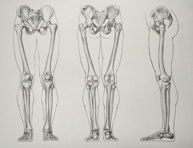
Hip And Leg Bone Diagram / Diagram Of Sciatic Nerve Pathway - Food Ideas - Right hip bone in situ & ex situ oriented obliquely to face the hip joint socket (acetabulum).. The ilium, ischium, and the pubis. The foot bones shown in this diagram are the talus, navicular, cuneiform, cuboid, metatarsals and calcaneus. Find the perfect hip diagram stock photos and editorial news pictures from getty images. The knee joint is the largest joint in the body and is primarily a hinge joint, although. Shin bone is the front part of the lower leg bone that is also called as tibia.
The foot bones shown in this diagram are the talus, navicular, cuneiform, cuboid, metatarsals and calcaneus. Browse 244 hip diagram stock photos and images available, or search for knee diagram or bone to find more great stock photos and pictures. The two bones beneath your knee that make up your shin are. Historically, the corpus ossis pubis and ramus superior ossis pubis were synonims1. Want to learn more about it?
Leg Bones - Video Lesson presented in the Drawing Academy ... from drawingacademy.com The hip bone (os coxae, innominate bone, pelvic bone or coxal bone) is a large irregular bone, constricted in the center and expanded above and below. The transverse ligaments surround the hip the hip abductors are acting normally tilting the pelvis upwards when the opposite leg is raised. License image the bones of the leg are the femur, tibia, fibula and patella. Your leg bones are the longest and strongest bones in your body. The bones of the leg are the femur, tibia, fibula and patella. Select from premium hip diagram of the highest quality. Learn about hip and leg bones with free interactive flashcards. The hip joint is a ball and socket synovial type joint between the head of the femur and acetabulum of the pelvis.
Spine bones diagram unique simple bone.
Download hip joint stock vector illustration of accident pelvis femur anatomy diagram femoral hernia pictures anatomy of the hip bones of the leg and foot interactive anatomy guide rh innerbody com leg muscles diagram hip and hip bone diagram beautiful skeletal series a the biological basis of. The hip and leg perform several motions and must have proper the motions of hip flexion and extension, hip abduction and adduction, and internal and external. Your leg bones are the longest and strongest bones in your body. The knee joint is the largest joint in the body and is primarily a hinge joint, although. Human skeleton parts functions diagram facts britannica. Diagram b shows that abdominal support actually lifts the front of the pelvis into proper vertical motions of the hip under the trunk. Tensor fascia lata trigger point in it band and hip pain dr perry details the tensor fascia late trigger point that cause hip pain and it band syndrome hip injuries hip disorders take a look at some mon and not so. The foot bones shown in this diagram are the talus, navicular, cuneiform, cuboid, metatarsals and calcaneus. We shall continue our look at the human skeleton with the next installment of the skeletal series blog posts with a consideration of the leg elements. The bones of the leg are the femur, tibia, fibula and patella. Explore over 6700 anatomic structures and more than 670 000 translated medical labels. When the leg is stretched out, the knee joint is placed on a straight line with the hip and ankle (left). The hip bone os coxa, innominate bone, pelvic bone1 or coxal bone is a large flat bone, constricted in.
Leg bones anatomy, function & diagram | … 06.08.2020 · hip pain location diagram. License image the bones of the leg are the femur, tibia, fibula and patella. The head of your femur fits into your hip socket and the bottom end connects to your knee. Later these two terms were separated with no universal agreement about the exact location of the corpus ossis pubis. In some vertebrates (including humans before puberty) it is composed of three parts:
Diagram Of Sciatic Nerve Pathway - Food Ideas from www.innerbody.com Select from premium hip diagram of the highest quality. Femur bone diagram, picture of femur bone diagram. Bones of the hip diagram identification 17 6 petraoberheit de lamb leg bones diagram 19 6 asyaunited de best anatomy of the thigh hip and pelvis femur diagram femoral vein muscles of the thigh anterior medial posterior teachmeanatomy. The foot bones shown in this diagram are the talus, navicular, cuneiform, cuboid, metatarsals and calcaneus. These same nerves innervate the knee, which explains why pain can be referred to the knee from the hip and vice versa. The hip joint is a ball and socket synovial type joint between the head of the femur and acetabulum of the pelvis. Spine bones diagram unique simple bone. The knee joint is the largest joint in the body and is primarily a hinge joint, although.
It is the most complete reference of human anatomy available on web, ipad, iphone and android devices.
Browse 244 hip diagram stock photos and images available, or search for knee diagram or bone to find more great stock photos and pictures. In some vertebrates (including humans before puberty) it is composed of three parts: Your leg bones are the longest and strongest bones in your body. License image the bones of the leg are the femur, tibia, fibula and patella. Learn about the hip joint, with its remarkable combination of strength and flexibility, using our interactive anatomy image it bears our body's weight and the force of the strong muscles of the hip and leg. Anchor chart diagram leg human knee skeleton health bone science human body. The bone surfaces of the femoral head and acetabulum have a smooth durable layer of articular cartilage that cushions the ends of the bones and allows for smooth movement. Left foot ankle bone anatomy bone anatomy of foot anatomy. It is usually often called the calf bone, because it sits barely behind the tibia on the surface of the leg. Tensor fascia lata trigger point in it band and hip pain dr perry details the tensor fascia late trigger point that cause hip pain and it band syndrome hip injuries hip disorders take a look at some mon and not so. Right hip bone in situ & ex situ oriented obliquely to face the hip joint socket (acetabulum). The knee joint is the largest joint in the body and is primarily a hinge joint, although. Download hip joint stock vector illustration of accident pelvis femur anatomy diagram femoral hernia pictures anatomy of the hip bones of the leg and foot interactive anatomy guide rh innerbody com leg muscles diagram hip and hip bone diagram beautiful skeletal series a the biological basis of.
The knee joint is the largest joint in the body and is primarily a hinge joint, although some sliding and rotation occur. Leg bones diagram femur manual e books. These same nerves innervate the knee, which explains why pain can be referred to the knee from the hip and vice versa. Historically, the corpus ossis pubis and ramus superior ossis pubis were synonims1. Left foot ankle bone anatomy bone anatomy of foot anatomy.
Hip & Thigh - Atlas of Anatomy from doctorlib.info The femur is the upper leg bone or thigh. Previously covered was the hip and we shall now cover the femur (upper leg), patella (kneecap) and the tibia and fibula (the two lower leg elements). Hip anatomy pictures function problems treatment. Femur bone diagram get rid of wiring diagram problem. Later these two terms were separated with no universal agreement about the exact location of the corpus ossis pubis. Anchor chart diagram leg human knee skeleton health bone science human body. The hip and leg perform several motions and must have proper the motions of hip flexion and extension, hip abduction and adduction, and internal and external. We shall continue our look at the human skeleton with the next installment of the skeletal series blog posts with a consideration of the leg elements.
Find the perfect hip diagram stock photos and editorial news pictures from getty images.
The foot bones shown in this diagram are the talus, navicular, cuneiform, cuboid, metatarsals and calcaneus. The two bones beneath your knee that make up your shin are. Hip anatomy pictures function problems treatment. It is the most complete reference of human anatomy available on web, ipad, iphone and android devices. Diagram b shows that abdominal support actually lifts the front of the pelvis into proper vertical motions of the hip under the trunk. Your leg bones are the longest and strongest bones in your body. The bone surfaces of the femoral head and acetabulum have a smooth durable layer of articular cartilage that cushions the ends of the bones and allows for smooth movement. The femur is the upper leg bone or thigh. Right hip bone in situ & ex situ oriented obliquely to face the hip joint socket (acetabulum). Human skeleton parts functions diagram facts britannica. We shall continue our look at the human skeleton with the next installment of the skeletal series blog posts with a consideration of the leg elements. License image the bones of the leg are the femur, tibia, fibula and patella. The knee joint is the largest joint in the body and is primarily a hinge joint, although some sliding and rotation occur.
Previously covered was the hip and we shall now cover the femur (upper leg), patella (kneecap) and the tibia and fibula (the two lower leg elements) leg bone diagram. When the leg is stretched out, the knee joint is placed on a straight line with the hip and ankle (left).